Scanning Electron Microscopy (SEM)
The Electron Microscopy facility at the James Hutton utilises two Carl Zeiss Scanning Electron Microscopes (SEM), complimented by multiple Bruker Energy Dispersive Spectroscopy (EDS) detectors, to provide high-resolution imaging and elemental analysis.
SEM/EDS aids in the analysis of samples to highlight topography, composition, and contamination, as well as characterising minerals and materials of unknown origin.
SEM/EDS – the power of precision
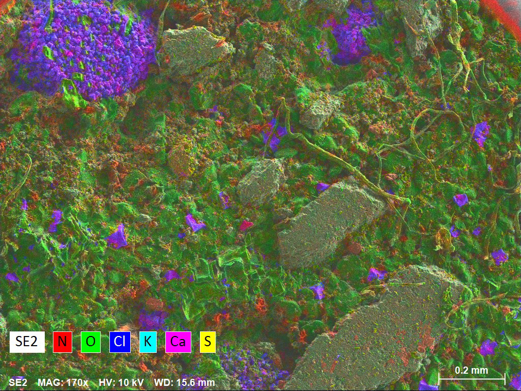
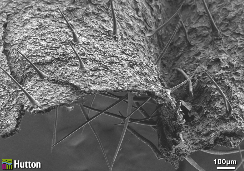
Scanning Electron Microscopy (SEM) provides high resolution images of sample topography using secondary electron imaging (SE), as well as highlighting compositional differences using back scattered electron imaging (BSE). Energy Dispersive Spectroscopy (EDS) allows elemental analysis, in addition to imaging, to aid the characterisation of samples. EDS can also be used to perform X-ray mapping, producing elemental maps, which visibly highlight by means of coloured maps, the elemental distribution within a sample.
These capabilities allow for the analysis of many different sample types, from a variety of industries from engineering to food. SEM/EDS provides valuable information in cases of unknown samples/contaminants, with visualisation of the material producing information on grain size and shape, as well as the associated elemental composition. This can play an important role in cases where unexpected changes have occurred and by performing a comparative analysis between a control and contaminated sample, can highlight the presence of micron sized deposits or damage. SEM/EDS is a non-destructive technique, which can be important where material or component parts are required to be returned or put for additional testing.
Allow the quality equipment and expertise within the Electron Microscopy department at the James Hutton Institute provide detailed, reliable analysis to help optimize processes, enhance quality and problem solve within your industry.
Laura-Jane Strachan, Head of Electron Microscopy
X-ray mapping can also be used in studies into corrosion of various material types, where failure has been caused by chemical and/or physical changes to the material in question.
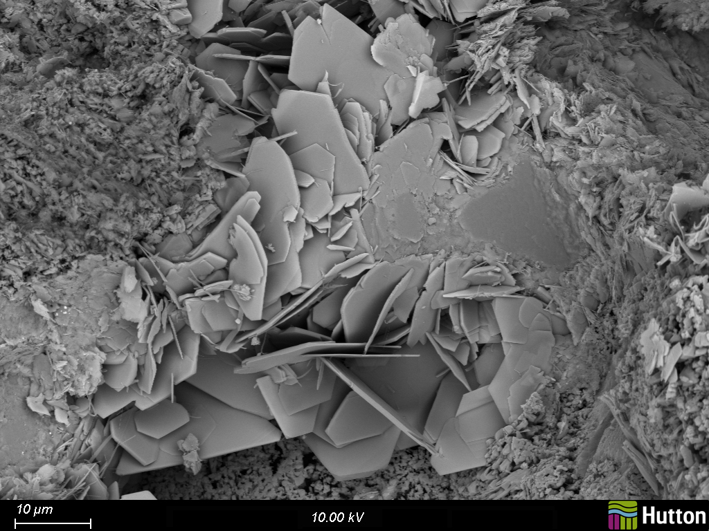
SEM/EDS is commonly used for Minerology investigations, including the characterisation of rock samples to provide a detailed description of mineral types, grain size and shape, potential origin (detrital or diagenetic), morphology, distribution and their inter-relationships. It is the only technique which allows the visualisation of the minerals present, and therefore plays an important role in characterising these types of samples.
SEM/EDS is also valuable in the study of scales. Information on the structure and the elemental composition of the particles, or layers in cases of larger scales, obtained by SEM/EDS analysis can provide vital information to customers as to the effectiveness of treatments or plans for further treatments.


Contact for more information
Laura-Jane Strachan
Head of Electron Microscopy
Based in Aberdeen
Laura-Jane Strachan gained a degree in Geology and an honors degree in Geography from the University of Aberdeen. She came to the James Hutton Institute in 2011 with over 5 years experience of working in the oil and gas industry. Her previous experience is in the identification of solid and fluid induced mechanisms causing formation damage, using standard and cryogenic SEM methods and thin section analysis
Laura-Jane currently works in the Electron Microscopy section and predominantly works on commercial projects, using Scanning Electron Microscopy to characterise a range of sample types.
